
Book Appointment Now
Digital Planning and Three-Dimensional Surgical Evaluation
Digital planning and three-dimensional surgical assessment is an important stage that allows treatment to be structured correctly from the very beginning, especially in patients with advanced bone deficiency. In implant or bone graft procedures performed without digital planning and three-dimensional surgical assessment, the physician’s experience is of course highly valuable; however, it is not sufficient on its own. This is because it is not possible to fully see the real volume of the jawbone, the position of the nerves, the location of the sinus cavities and the suitable areas for implants through visual examination alone.
In some patients, tooth loss may have occurred many years ago. During this period, the jawbone may have gradually become thinner and lost its height or width. Most of the time, the patient is not aware of this. From the outside, only the missing tooth is visible; however, when implant treatment is planned, the real issue is whether the bone can support the implant. This is where three-dimensional imaging and digital planning come into play.
Today, in modern implant surgery, it has become much more possible to see, measure and analyze the patient’s oral structure in detail before starting treatment. When panoramic X-rays, three-dimensional dental tomography and intraoral digital scanners are used together, a very clear picture is created for the physician. Thanks to this picture, the question is not only “Can an implant be placed?”; the location, angle, depth and bone support of the implant are also planned in advance.
İçindekiler Tablosu - Table of Contents
- 1 Why Are Digital Planning and Three-Dimensional Surgical Assessment So Important?
- 2 Which Imaging Methods Are Used in Digital Planning and Three-Dimensional Surgical Assessment?
- 3 Treatment of Advanced Bone Deficiency with Digital Planning and Three-Dimensional Surgical Assessment
- 4 Iliac Bone Grafting with Digital Planning and Three-Dimensional Surgical Assessment
- 5 Implant Planning with Digital Planning and Three-Dimensional Surgical Assessment
- 6 What Do Digital Planning and Three-Dimensional Surgical Assessment Mean for the Patient?
- 7 In Which Patients Do Digital Planning and Three-Dimensional Surgical Assessment Become More Critical?
- 8 How Do Digital Planning and Three-Dimensional Surgical Assessment Affect the Treatment Outcome?
- 9 Frequently Asked Questions
- 9.1 Which Stages Do Digital Planning and Three-Dimensional Surgical Assessment Include?
- 9.2 In Which Situations Are Digital Planning and Three-Dimensional Surgical Assessment Especially Necessary?
- 9.3 Are Digital Planning and Three-Dimensional Surgical Assessment Painful Procedures?
- 9.4 Can Implants Be Placed Immediately After Digital Planning and Three-Dimensional Surgical Assessment?
- 9.5 Where Are Digital Planning and Three-Dimensional Surgical Assessment Performed?
Why Are Digital Planning and Three-Dimensional Surgical Assessment So Important?
In patients with advanced bone loss, treatment is often more complex than a standard implant procedure. This is because there may not be sufficient bone volume in the area where the implant will be placed. Sometimes the bone height may appear sufficient, but the width may be inadequate. Sometimes the nerve canal or sinus cavity may directly affect implant planning. These details cannot always be understood clearly enough with two-dimensional images.
Digital planning and three-dimensional surgical assessment give the physician the opportunity to examine the patient’s jaw structure in three dimensions. Thus, the bone is evaluated not only from top to bottom, but also from front to back and from right to left. This approach is especially valuable before advanced surgical procedures such as iliac bone grafting. Because the amount of missing bone, the area where bone needs to be added and how the implants will be placed afterward can be calculated before treatment even begins.
From the patient’s perspective, this planning process also provides reassurance. Many people naturally feel anxious before a surgical procedure. They want to know how the treatment will progress, how many stages it will involve and why certain additional procedures may be needed. Explanations made through digital images help the patient understand the treatment process better. This also strengthens communication between the patient and the physician.
Which Imaging Methods Are Used in Digital Planning and Three-Dimensional Surgical Assessment?
One of the first stages in treatment planning is a panoramic X-ray. A panoramic X-ray provides a general view of the oral and jaw structure. The condition of existing teeth, missing teeth, general bone level, impacted teeth or obvious pathological conditions can be evaluated through this image. However, since panoramic X-rays are two-dimensional, they provide limited information about bone thickness and depth.
For a more detailed assessment, three-dimensional dental tomography, in other words CBCT, is used. This imaging method is very important for seeing the real volume of the jawbone. Bone height, bone width, the position of the nerves, sinus cavities and anatomical structures can be examined millimetrically. Especially in patients planned for implant surgery or bone grafting, three-dimensional tomography becomes one of the main guides of treatment.
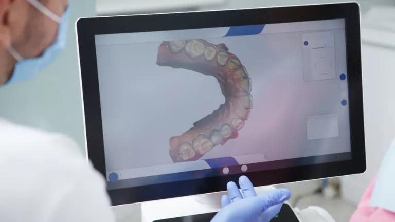
Intraoral digital scanners record the patient’s teeth, gums and bite structure in three dimensions. Compared to traditional impression methods, they offer a more comfortable process for the patient. When digital scan data is combined with tomography images, both the bone structure and the intraoral surfaces can be evaluated in the same digital environment. This combination contributes to more realistic surgical planning.
Treatment of Advanced Bone Deficiency with Digital Planning and Three-Dimensional Surgical Assessment
Advanced bone deficiency is one of the most important conditions to consider in implant treatment. Long-term toothlessness, infections, trauma, unsuccessful tooth extractions or previous surgical procedures may cause bone loss in the jawbone. This loss may sometimes be mild and can be managed with minor support procedures. However, in some patients, more comprehensive bone reconstruction may be required before implants can be placed.
Digital planning and three-dimensional surgical assessment make the treatment plan healthier by clarifying the extent of bone deficiency. For example, if there is horizontal bone loss only in one area, a different surgical approach may be required. If both height and width loss are present, more advanced bone grafting procedures may come into consideration. In areas close to the sinus region in the upper jaw, the sinus anatomy must also be evaluated separately.
At this point, the goal is not only to replace the missing bone. The main objective is to create a bone foundation on which future implants can function healthily for many years. Therefore, bone grafting and implant planning should not be considered separately. Proper digital planning affects not only today’s surgical stage of treatment, but also the future prosthetic stage.
Iliac Bone Grafting with Digital Planning and Three-Dimensional Surgical Assessment
Iliac bone grafting is one of the important surgical methods used in patients with advanced bone deficiency. Bone tissue taken from the iliac region is applied to the deficient area in the jawbone to help create new bone volume. This procedure is generally preferred in more advanced cases where standard bone powder applications would not be sufficient.
Planning before such a surgical procedure must be carried out very carefully. This is because the borders of the area where the graft will be applied, the amount of bone needed and the future position of the implants must be determined in advance. Digital planning and three-dimensional surgical assessment allow this process to progress in a more controlled manner. The physician can see more clearly where the missing bone begins, in which direction and to what extent volume loss has occurred, and how the implants can be positioned after graft application .
This approach both increases surgical predictability and makes the stages of the treatment process more understandable. In patients who will undergo iliac bone grafting, not only the existing deficiency but also the functional and aesthetic result aimed for at the end of treatment must be considered. Because a successful result depends not only on replacing the bone, but also on placing the implants in the correct position.
Implant Planning with Digital Planning and Three-Dimensional Surgical Assessment
One of the most common mistakes in implant treatment is focusing only on the area where the bone seems suitable. However, the correct position of the implant is important not only for its integration with the bone, but also for the natural appearance of the tooth to be placed on it and for balanced distribution of chewing forces. Therefore, implant planning should be carried out with both surgical and prosthetic perspectives.
Digital planning and three-dimensional surgical assessment make it possible to evaluate many factors simultaneously while determining the ideal implant position. Bone volume, distance from neighboring teeth, bite relationship with the opposing jaw and aesthetic expectations are considered together. This planning is much more sensitive, especially for implants placed in the anterior region. Because even a difference of a few millimeters in the front tooth area can affect the appearance of the gums and smile aesthetics.
In the posterior regions, chewing forces are greater, so placing the implant at the correct angle becomes even more important. In implant applications to be performed near the nerve canal in the lower jaw or near the sinus cavity in the upper jaw, making a decision without three-dimensional imaging would not be appropriate. For this reason, digital planning in implant surgery is no longer a luxury application today; it is an important necessity for the safe progress of treatment.
What Do Digital Planning and Three-Dimensional Surgical Assessment Mean for the Patient?
Patients may often think that digital planning is only a technical detail that helps the physician. However, this process also directly affects the patient’s treatment experience. First of all, the patient can see their own oral and jaw structure in a more understandable way. Where the bone deficiency is, why the implant cannot be placed immediately or why a bone graft is needed can be explained more clearly through digital images.
This clarity increases trust in the treatment. Because the patient does not only hear what is said, but also understands their own condition by seeing it. Especially in people with advanced bone loss, treatment may consist of several stages. In such a process, knowing what to expect can also be psychologically comforting for the patient.
Digital planning also contributes to a more organized surgical procedure. Detailed preparation before the operation helps decisions to be made more clearly during the procedure. This positively affects both the physician’s working comfort and the patient’s sense of trust throughout the treatment process.
In Which Patients Do Digital Planning and Three-Dimensional Surgical Assessment Become More Critical?
Digital planning is beneficial for many patients considering implant treatment; however, in some cases, it becomes much more critical. Patients who have been toothless for many years, those with advanced bone resorption, people who have previously experienced implant loss, patients planned for bone grafting and individuals who will receive implants in areas close to the sinus or nerve canal are included in this group.
Three-dimensional assessment is also important in full-arch implant applications, multiple tooth deficiencies and cases requiring advanced jaw surgery. Because in such treatments, not a single implant but the entire oral structure must be planned in a balanced way. The distance between implants, their angles, prosthetic design and distribution of chewing forces affect long-term success.
In our clinic in Galata, Beyoğlu, Istanbul, advanced bone deficiency treatments, iliac bone graft operations, implant surgery and advanced jaw surgery procedures are evaluated with digital planning technologies. A personalized treatment approach is created for each patient using three-dimensional dental tomography, intraoral digital scanning and computer-assisted surgical planning systems.
How Do Digital Planning and Three-Dimensional Surgical Assessment Affect the Treatment Outcome?
A successful implant treatment is not measured only by the implant’s integration with the bone. The implant must be in the correct position, the tooth placed on it must look natural, chewing forces must be carried in a balanced way and the patient must be able to use it comfortably in the long term. Therefore, the treatment outcome is directly related to the decisions made during the planning stage.
Thanks to digital planning and three-dimensional surgical assessment, possible difficulties can be seen more clearly before treatment begins. If bone volume is insufficient, grafting is planned. If the sinus cavity is close, the surgical approach is determined accordingly. In areas close to the nerve canal, implant length and angle are carefully evaluated. In the aesthetic zone, gum level and the future appearance of the prosthesis are taken into account.
All these details contribute to a more controlled treatment process. Instead of a standard plan that does not suit the patient’s oral structure, a personalized roadmap is created. This difference is felt much more clearly, especially in cases with advanced bone deficiency. Because bone loss does not develop in the same way in every patient, and every treatment does not progress in the same order.
Frequently Asked Questions
Which Stages Do Digital Planning and Three-Dimensional Surgical Assessment Include?
- First, an intraoral examination is performed and the general treatment need is evaluated.
- The jaw structure and the general condition of existing teeth are examined with a panoramic X-ray.
- Bone height, width and anatomical structures are analyzed with three-dimensional dental tomography.
- The teeth and gum structure are recorded in three dimensions with an intraoral digital scanner.
- Tomography and digital scan data are combined in planning software.
- The position of the implants, the need for bone grafting and the surgical approach are planned individually.
This process allows treatment to progress more clearly and in a more controlled manner, especially in patients with advanced bone deficiency. Thus, before the operation, not only the current condition but also the targeted result at the end of treatment is evaluated.
In Which Situations Are Digital Planning and Three-Dimensional Surgical Assessment Especially Necessary?
- In patients with advanced bone loss
- In cases planned for iliac bone grafting
- In upper jaw implants close to the sinus region
- In implant applications adjacent to the nerve canal in the lower jaw
- In cases where implant loss has occurred before
- In full-arch implant treatments
- In aesthetic zone implant applications
- In patients requiring complex jaw surgery
In these situations, digital planning should be considered not only as a technology that facilitates treatment, but also as a fundamental assessment method that enables the right decisions to be made.
Are Digital Planning and Three-Dimensional Surgical Assessment Painful Procedures?
The imaging and scanning procedures used in the digital planning process are not painful. Panoramic X-rays and three-dimensional dental tomography are imaging procedures completed in a short time. Intraoral digital scanning is more comfortable for many patients compared to traditional impression methods. No surgical procedure is performed at this stage; only the necessary data for treatment planning is collected.
Can Implants Be Placed Immediately After Digital Planning and Three-Dimensional Surgical Assessment?
This depends on the patient’s bone structure. If there is sufficient bone volume for an implant, implant surgery may be planned if the physician considers it appropriate. However, in patients with advanced bone deficiency, bone grafting or bone reconstruction may first be required. After advanced surgical procedures such as iliac bone grafting, it may be necessary to wait for the appropriate healing period before implant placement. Therefore, the process is evaluated individually for each patient.
Where Are Digital Planning and Three-Dimensional Surgical Assessment Performed?
Digital planning and three-dimensional surgical assessment are performed in clinics that have the necessary imaging systems and surgical planning infrastructure. In our clinic in Galata, Beyoğlu, Istanbul, advanced bone deficiency treatments, iliac bone graft operations, implant surgery and advanced jaw surgery procedures are evaluated with three-dimensional dental tomography, digital intraoral scanning systems and computer-assisted surgical planning technologies. In this way, the treatment process is planned individually according to the patient’s existing bone structure, aesthetic expectations and long-term functional needs.